How to Identify and Treat Feline Dental Resorptive Lesions
You’ll want to spot feline tooth resorption early-look for drooling, jaw chattering, or your cat avoiding dry food, since 67% of cats develop this painful condition. Half of lesions hide below the gumline, so annual dental X-rays under anesthesia are essential. Once confirmed, treatment depends on type: Type 1 needs full extraction, while Type 2 may allow crown amputation if ankylosis is present. Always use dental explorers and radiographs for accurate diagnosis. Post-op care includes nerve blocks and multi-day pain relief, ensuring your cat recovers comfortably. There’s more to learn about keeping their mouth healthy and pain-free.
We are supported by our audience. When you purchase through links on our site, we may earn an affiliate commission, at no extra cost for you. Learn more. Last update on 19th July 2026 / Images from Amazon Product Advertising API.
Notable Insights
- Feline dental resorptive lesions affect up to 67% of cats and often start below the gumline at the mandibular third premolar.
- Nearly 50% of lesions are invisible without dental radiographs, making full-mouth X-rays under anesthesia essential for accurate diagnosis.
- Type 1 lesions have normal root density and require complete extraction; crown amputation is not safe.
- Type 2 lesions involve root resorption and dentoalveolar ankylosis, allowing crown amputation if no periapical disease is present.
- Annual veterinary dental exams with radiographs and at-home monitoring for eating changes help detect and manage resorptive lesions early.
What Is Feline Tooth Resorption? (And Why It’s So Painful)
Ever wonder why your cat suddenly flinches while eating or drools more than usual? You’re likely seeing signs of feline odontoclastic resorptive lesions (FORLs), a common but painful condition affecting up to 67% of cats. Resorption starts when odontoclast cells attack the tooth, often beginning subgingivally at the cementum near the mandibular third premolar. As destruction progresses into dentin and reaches the pulp cavity, pain increases-though your cat may hide it. Even subtle jaw chattering or hesitance with dry food can signal trouble. Resorption isn’t just surface wear; it’s active tooth loss from within. Left untreated, it causes chronic pain, eating issues, and risks like hepatic lipidosis. Radiographic staging (Stage 1–5) helps determine severity, guiding care. Knowing this? You’re already ahead in protecting their comfort and health.
How Diagnosis Works: Spotting Resorption With X-Rays
You can’t rely on what you see in your cat’s mouth alone-nearly half of all feline resorptive lesions are invisible during a surface exam, which is why dental radiographs are the gold standard for diagnosis. Radiographic evidence reveals subgingival lesions and early lesions before structural damage is visible. Full-mouth radiographic surveys under anesthesia allow complete radiographic evaluation, critical for accurate diagnosis and treatment. These surveys detect feline tooth resorption that might otherwise be missed, and distinguish between Type 1 and type 2 resorption patterns. You’ll need radiographic evaluation to identify dentoalveolar ankylosis and rule out periapical rarefaction before any crown amputation.
| Finding | Seen on Radiograph? | Clinical Sign? |
|---|---|---|
| Early lesions | Yes | No |
| Subgingival resorption | Yes | No |
| Type 1 and type 2 | Yes | Sometimes |
| Dentoalveolar ankylosis | Yes | No |
| Full-mouth involvement | Yes | Variable |
Type 1 Vs Type 2: What the Results Mean for Treatment
Dental X-rays don’t lie-when it comes to feline tooth resorption, they reveal the real story beneath the gumline, and that’s where the difference between Type 1 and Type 2 lesions becomes critical. You’ll see that type 1 and type 2 root resorption differ markedly on dental radiographic images. Type 1 lesions show normal root density with inflammation often tied to periodontal disease, requiring full extraction-crown amputation with intentional root retention isn’t safe here. Leaving an orphaned root can worsen advanced feline resorptive issues. Type 2, commonly linked to metabolic factors, shows replacement resorption and dentoalveolar ankylosis; Harvey CE noted it allows crown amputation with intentional root retention as a valid option. Misreading clinical signs may lead to improper treatment, so always confirm lesion type. Proper imaging guides safe decisions, ensuring your cat avoids unnecessary pain or complications from mismanaged feline dental resorptive lesions.
Feline Tooth Resorption Treatment: Extraction and Care
A proper extraction starts with a clear picture-literally-so dental radiographs under anesthesia are non-negotiable when treating feline tooth resorption. You’ll need general anesthesia to perform dental radiographs and accurately assess root structure, especially to distinguish between Type 1 and Type 2 resorption. For Type 1, you must do a complete extraction of crown and root; any remnant can cause pain and infection. Type 2 may allow crown amputation with intentional root retention, but only if radiographs confirm full dentoalveolar ankylosis and no periapical disease. Oral surgery techniques like gingival flaps, bone removal, or sectioning help achieve clean extractions. Always use preoperative nerve blocks and follow with multi-day analgesia as part of postoperative pain management-these cats often have chronic discomfort. Your goal is thoroughness and long-term comfort.
Prevent Pain: At-Home Care Tips and Checkups
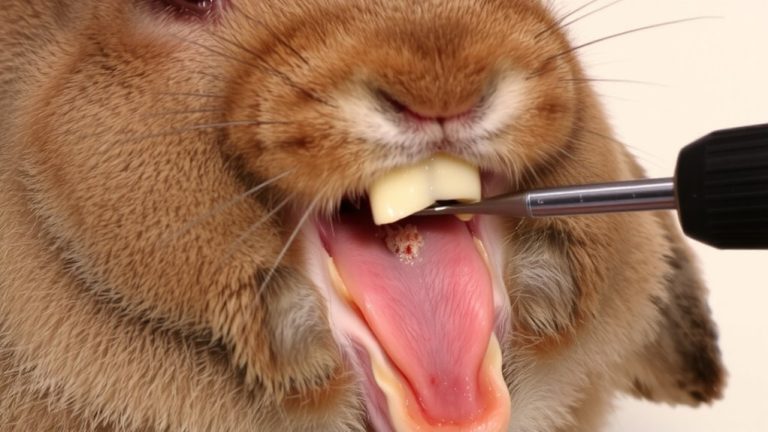
While tooth resorption often progresses silently, staying ahead of pain means combining routine at-home observations with professional care, starting with annual veterinary dental checkups that include full-mouth radiographs-because 53% of cats have visible lesions and nearly half show damage only detectable on X-rays. During each oral examination, your vet uses a dental explorer to probe for defects and relies on radiographic imaging to uncover hidden disease. At home, watch for clinical signs like jaw chattering, drooling, or avoiding dry kibble, since early detection supports better pain management. Brush your cat’s teeth with veterinary-approved toothpaste to reduce plaque, though it won’t stop feline tooth resorption. Feed a balanced diet avoiding excess vitamin D, as imbalances may contribute to lesions. Regular at-home care and annual checkups are key pillars in protecting your cat’s comfort and long-term oral health.
On a final note
You’ve got this-catching tooth resorption early means less pain for your cat. Always check for drooling, jaw chattering, or favoring one side when eating. Dental X-rays are key, since lesions hide below the gumline. If your vet finds Type 1 or Type 2 resorption, extraction is the gold standard. Post-op, stick to soft food for 7–10 days. Use chlorhexidine gel daily, brush with PetMD toothpaste, and schedule cleanings every 6 months to stay ahead.