Feline Corneal Sequestrum: Symptoms, Diagnosis & Treatment
You’ll notice a dark brown or black spot on your cat’s eye, often with tearing, squinting, or pawing-common in Persians and Himalayans due to herpesvirus or dry eye. Vets use fluorescein stain and slit-lamp exams to confirm it. Surgery, like lamellar keratectomy, usually removes the lesion, sometimes with a conjunctival graft. Post-op care includes antibiotics, antivirals, lubricants, and an E-collar for 2–4 weeks. Healing takes 4–6 weeks, but recurrence reaches 30%, so ongoing eye checks and antiviral support are key. Watch for subtle signs early-they could save your cat’s vision.
We are supported by our audience. When you purchase through links on our site, we may earn an affiliate commission, at no extra cost for you. Learn more. Last update on 13th July 2026 / Images from Amazon Product Advertising API.
Notable Insights
- Look for a dark brown or black spot on the cornea, often with tearing, squinting, or pawing at the eye.
- Confirm diagnosis using fluorescein staining, slit-lamp exam, and Schirmer tear test to assess ulcers and tear production.
- Most cases require surgical removal via lamellar keratectomy, especially for deep or painful sequestra.
- Post-surgery, use topical antibiotics, antivirals, lubricants, and an Elizabethan collar to prevent complications.
- Monitor for recurrence, especially in brachycephalic breeds, and manage underlying conditions like dry eye or herpesvirus.
What Is Feline Corneal Sequestrum?
Think of it as a dark spot on your cat’s eye that just won’t go away-that’s feline corneal sequestrum. This condition, unique to cats, appears as a dark brown spot caused by necrotic corneal tissue dying off in the central or paracentral cornea. You’re most likely to see corneal sequestrum in cats with chronic corneal irritation, often tied to feline herpesvirus or corneal ulcers. It’s not just surface-level damage-this dead tissue can go deep, weakening the corneal structure over time. Brachycephalic breeds, like Persians and Himalayans, are especially prone due to their facial anatomy. Unlike other eye issues, corneal sequestra form gradually and persist without treatment. The affected corneal tissue becomes pigmented and non-viable, creating a risk for ulceration or even perforation. Early detection matters-this isn’t something that resolves on its own.
How Do You Spot the Symptoms Early?
What if that faint dark speck in your cat’s eye isn’t just a harmless mark? It could be an early sign of corneal sequestrum, a condition where dark pigmentation forms on the cornea, often starting as a small brown or black spot. In cats, early symptoms include subtle squinting (blepharospasm), excessive tearing (epiphora), or pawing at the eye. You might also notice vascularization-tiny blood vessels spreading from the eye’s edge toward the center. Even mild discomfort, light sensitivity, or third eyelid elevation can signal trouble. Brachycephalic breeds like Persians and Himalayans are especially prone, so routine eye checks help catch sequestrum before deep ulceration occurs. Don’t assume discoloration is just pigmentation-early detection means simpler treatment and better outcomes for your cat.
Why Are Some Cats More at Risk for Feline Corneal Sequestrum?
That faint dark spot you noticed in your cat’s eye isn’t just a random mark-it could signal the start of corneal sequestrum, especially if your cat belongs to a high-risk breed. You see, brachycephalic cats like Persian and Himalayan breeds are more predisposed due to flatter faces that cause incomplete eyelid closure, increasing cornea exposure. These cats often have reduced tear production-sometimes up to 30% lower-raising the risk of corneal damage. Conditions like entropion, where eyelids roll inward, further irritate the cornea. Plus, feline herpesvirus (FHV-1) lurks in many brachycephalic cats, with over 80% of corneal sequestra linked to active viral infection. Genetic factors make certain feline breeds up to five times more likely to develop this condition. Keeping an eye on tear film stability and signs of eye discomfort in predisposed breeds is essential for early care.
How Do Vets Diagnose Feline Corneal Sequestrum?
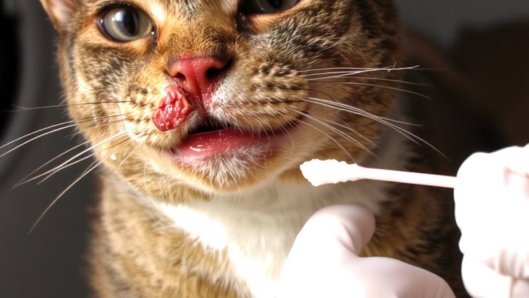
How does your vet pinpoint that dark spot in your cat’s eye as a corneal sequestrum and not something else? Through a thorough ophthalmic examination, they identify the classic signs of feline corneal sequestrum-a dark brown to black lesion often surrounded by vascularization and irritation. Your vet will use fluorescein staining to reveal any underlying corneal ulcer, which glows green under cobalt blue light. Slit-lamp biomicroscopy gives a magnified view, evaluating how deep the sequestrum has infiltrated the corneal stroma. They’ll also perform a Schirmer tear test to check for keratoconjunctivitis sicca, especially common in Persians. Tonometry measures intraocular pressure to rule out secondary glaucoma. These steps-fluorescein staining, slit-lamp biomicroscopy, Schirmer tear test, and tonometry-ensure an accurate diagnosis and help guide your cat’s recovery.
Is Surgery Necessary for Feline Corneal Sequestrum?
Once your vet confirms a corneal sequestrum through fluorescein staining, slit-lamp evaluation, and other diagnostic steps, the next question is clear: surgery is usually the best path forward, especially for anything beyond a very shallow lesion. For most feline corneal sequestra, surgical intervention is necessary-lamellar keratectomy is the gold standard to remove necrotic tissue and prevent corneal perforation. Deep sequestra, which extend into the stroma, almost always require this procedure under general anesthesia. A veterinary ophthalmologist often performs the surgery and may place a conjunctival graft afterward to support healing and reduce recurrence. While rare, superficial cases might be managed medically, but excision is preferred to relieve pain and avoid complications. Without surgery, risks like secondary infection, chronic discomfort, and potential eye loss rise markedly-especially with ulceration or herpesvirus involvement.
What to Expect After Feline Corneal Sequestrum Removal
After your cat’s corneal sequestrum removal, you’ll need to keep an Elizabethan collar on at all times for at least 2 to 3 weeks-most vets recommend 4-to prevent scratching or rubbing that could disrupt healing. Postoperative care includes pain management, as inflammation may temporarily worsen discomfort before improving with prescribed meds. You’ll apply topical antibiotics, antiviral medications (especially if herpesvirus is involved), and ocular lubricants several times daily. Short-term atropine helps with pupil dilation to reduce ciliary spasm pain but is discontinued early to avoid tear film issues. Healing time typically ranges from 4 to 6 weeks. Your veterinary ophthalmologist will schedule rechecks to monitor progress, guarantee graft integration, and watch for early signs of corneal sequestrum recurrence.
Can Feline Corneal Sequestrum Come Back?
Could your cat’s corneal sequestrum come back? Yes, recurrence happens in up to 30% of cases, especially in breeds like Persian and Himalayan cats. Even after successful surgical removal, the condition can return in the same eye or affect the other one, often tied to unresolved feline herpesvirus. If underlying causes-like corneal dryness or eyelid issues-are left untreated, risk increases. Complete recovery depends on managing these factors long-term. Your vet may prescribe antiviral therapy and daily artificial tears to keep the surface moist and suppress viral flare-ups. Regular ophthalmic recheck exams are essential; they catch early signs of recurrence before serious damage occurs. Staying consistent with treatment and follow-ups gives your cat the best chance at lasting comfort and clear vision. Don’t skip checkups-even if eyes look healed, silent issues can linger.
On a final note
You’ll spot feline corneal sequestrum as a dark spot on your cat’s eye, often with tearing or squinting. Flat-faced breeds like Persians are most at risk. Vets diagnose it with a fluorescein test, and surgery-like a keratectomy-is usually needed. After removal, use prescribed ointments, like Terramycin, twice daily. Most cats heal well in 2–3 weeks, but recurrence happens, so monitor closely and schedule follow-ups every 3 months.