Understanding Canine Neosporosis: Causes, Symptoms, and Diagnosis
You can catch canine neosporosis when your dog ingests Neospora caninum oocysts from contaminated soil, water, or raw beef with tissue cysts, often near infected dog or coyote feces. Watch for hind limb weakness, muscle atrophy, seizures, or megaesophagus causing dysphagia. Confirm infection with an IFAT titer ≥1:200 or positive PCR on CSF. Less than 6% fully recover from neurological signs, so early testing and vets-approved protocols make all the difference - especially if you act fast when symptoms start.
We are supported by our audience. When you purchase through links on our site, we may earn an affiliate commission, at no extra cost for you. Learn more. Last update on 19th July 2026 / Images from Amazon Product Advertising API.
Notable Insights
- Canine neosporosis is caused by the protozoan parasite Neospora caninum, transmitted via ingestion of oocysts from infected dog feces or tissue cysts in raw meat.
- Vertical transmission occurs from infected dams to puppies during pregnancy, a major route of infection in dogs.
- Neurological signs include hind limb paresis, muscle atrophy, seizures, and paralysis, often progressing rapidly over weeks.
- Megaesophagus may develop, causing dysphagia and increasing the risk of aspiration pneumonia in affected dogs.
- Diagnosis combines IFAT titers (≥1:200 indicates active infection) with PCR detection of N. caninum DNA in CSF or tissues for confirmation.
What Causes Canine Neosporosis?
While it might seem like something only seen in veterinary textbooks, canine neosporosis is a real and serious condition caused by a microscopic parasite called *Neospora caninum*. This obligate intracellular protozoan parasite spreads when your dog ingests sporulated oocysts from contaminated soil or water, often shed by definitive hosts like infected dogs or coyotes. Alternatively, they can consume tissue cysts in intermediate hosts-commonly cattle-containing bradyzoites. Once inside, the parasite transforms into tachyzoites, spreading systemically and damaging neuromuscular tissues. Vertical transmission is especially concerning, as transplacental infection during pregnancy can severely affect unborn puppies. Prenatal cases often result in stillbirth or weakness, even with proper nutrition and care. Though unsporulated oocysts are initially shed in feces, they mature into infective forms within 1–3 days outdoors. You can’t see them, but they persist, making prevention critical.
How Does Neospora Caninum Spread to Dogs?
How does your dog end up infected with *Neospora caninum*? Dogs pick up the infection mainly by ingesting sporulated oocysts from contaminated food, water, or soil-these oocysts come from the feces of infected definitive hosts like canids. You should also know your dog can get Neosporosis through horizontal transmission by eating raw or undercooked meat containing tissue cysts from intermediate hosts such as cattle or deer. Puppies often face vertical transmission, where the parasite passes from dam to offspring during pregnancy or via infected colostrum and milk. Unsporulated oocysts develop within 5–7 days post-infection and are shed in feces for up to 3 weeks.
| Transmission Route | Source of Infection |
|---|---|
| Ingesting sporulated oocysts | Contaminated feces |
| Horizontal transmission | Tissue cysts in meat |
| Vertical transmission | Infected dam to fetus |
| Postnatal infection | Infected milk/colostrum |
| Environmental exposure | Soil, water, food |
Clinical Signs of Canine Neosporosis
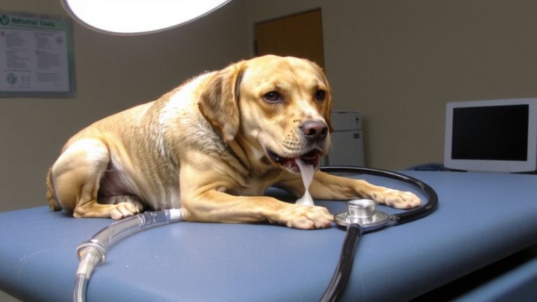
Your dog may show serious neurological symptoms if infected with *Neospora caninum*, especially when the parasite spreads during pregnancy or takes hold early in life. Common clinical signs include hind limb paresis, which often progresses to rigid paralysis and muscle atrophy, particularly in puppies under one year. You might notice your dog struggling to stand or walk, with stiffness worsening over days to weeks. Neurological signs like seizures, behavioral changes, and even blindness can occur, especially in adult dogs with multifocal CNS involvement. Other symptoms include dysphagia, often linked to megaesophagus, which increases aspiration risk. Cervical muscle weakness, incontinence, and skin ulcers may also appear. In severe cases, respiratory muscle paralysis can develop, especially with early, widespread tachyzoite dissemination. While rare, complete recovery occurs in under 6% of neurological cases, underscoring the poor prognosis tied to neuromuscular disease.
Confirming Neospora: Serology, PCR, and Clinical Signs
What does it take to confirm Neospora in your dog? You’ll need a mix of serology, PCR, and clinical signs to reach a solid diagnosis. An IFAT titer ≥1:200 in serum or CSF suggests active infection, while ≥1:50 means exposure. Pairing IFAT with PCR on CSF or tissue boosts sensitivity-some dogs with titers like 1:400 test positive for tachyzoites via PCR, even though 58.1% of seropositive dogs have negative PCR. Oocysts rarely show up in feces due to low, intermittent shedding, and must be DNA-confirmed to rule out Hammondia heydorni. For definitive diagnosis, PCR detection of N. caninum DNA in CSF, tissue, or feces is key-samples stay stable up to 10 days at 4°C. In puppies, clinical signs like hindlimb paresis, cervical weakness, or rigidity, plus cattle exposure or maternal abortion history, strengthen the case.
On a final note
You can protect your dog by understanding neosporosis, caused by *Neospora caninum*, often from eating infected raw meat or contaminated soil. Watch for muscle weakness, lameness, or neurological signs. Diagnosis combines PCR, serology, and clinical symptoms. Prevent exposure with balanced commercial diets-avoid raw feeding. Keep your yard clean, secure trash, and test promptly if symptoms appear. Early action means better outcomes, so stay informed and proactive about your pet’s health.